- SEO Powered Content & PR Distribution. Get Amplified Today.
- Platoblockchain. Web3 Metaverse Intelligence. Knowledge Amplified. Access Here.
- Source: https://www.nanowerk.com/nanotechnology-news2/newsid=62405.php
- 10
- 11
- 9
- a
- ability
- Able
- About
- accuracy
- Achieve
- and
- applicable
- Array
- average
- averaging
- based
- because
- BEST
- Better
- billions
- Bottom
- capable
- cases
- Center
- Changes
- chemical
- class
- closely
- closer
- Column
- complementary
- components
- could
- crowd
- Current
- data
- Date
- day
- decades
- demonstrating
- density
- Department
- depends
- detail
- detected
- determining
- developed
- Development
- Diamond
- different
- differentiate
- direction
- diverse
- down
- dynamics
- each
- effect
- efficiency
- effort
- electrons
- Equivalent
- especially
- essential
- essentially
- example
- existing
- field
- Football
- from
- function
- Furthermore
- future
- Germany
- getting
- giant
- Go
- goal
- going
- grant
- great
- groundbreaking
- Hopefully
- How
- HTTPS
- Hundreds
- image
- Imaging
- important
- in
- incredible
- individual
- information
- initial
- Institute
- instrumental
- investigate
- issues
- IT
- itself
- Know
- lead
- Lives
- location
- Lot
- Low
- Magnetic field
- Making
- managed
- many
- mapping
- materials
- measures
- measuring
- method
- methods
- Middle
- might
- molecular
- molecule
- more
- most
- MRI
- Nature
- Need
- New
- novel
- ONE
- optimal
- order
- packed
- particularly
- perhaps
- person
- Pharmaceutical
- pharmaceuticals
- Physics
- pivotal
- plato
- Plato Data Intelligence
- PlatoData
- Point
- Point of View
- precise
- present
- probably
- Problem
- procedures
- processes
- provide
- Quantum
- quantum materials
- question
- ready
- recent
- remain
- research
- researchers
- Resolution
- resolved
- resonance
- review
- revolutionize
- ROW
- says
- Scale
- scan
- sensitive
- single
- Sitting
- Size
- small
- smaller
- So
- some
- special
- spins
- stages
- Step
- structural
- structure
- Student
- Study
- such
- synthetic
- Take
- techniques
- Technology
- ten
- The
- their
- Third
- times
- to
- tool
- top
- torn
- Trillion
- Trinity
- underlying
- understand
- university
- use
- various
- Vast
- View
- wanted
- Water
- What
- whether
- which
- while
- will
- Work
- would
- Your
- zephyrnet
More from Nanowerk

Development of cost-effective and strong composite carbon fiber using carbon nanotubes
Source Node: 1992517
Time Stamp: Mar 3, 2023

Microbial cell factories offer sustainable alternative to chemical factories for compound production
Source Node: 2375299
Time Stamp: Nov 9, 2023

Researchers identify two of the Milky Way’s earliest building blocks
Source Node: 2524560
Time Stamp: Mar 22, 2024


Imaging agents light up two cancer biomarkers at once to give more complete picture of tumor
Source Node: 2121965
Time Stamp: Jun 5, 2023
New technique unlocks tunable plasmonics in promising photonic glass
Source Node: 2415845
Time Stamp: Dec 27, 2023

New X-ray imaging technique to study the transient phases of quantum materials
Source Node: 1850118
Time Stamp: Dec 22, 2022

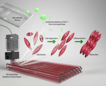
New and improved bioink to enhance 3D bioprinted skeletal muscle constructs
Source Node: 2241853
Time Stamp: Aug 29, 2023
3D-printed plasmonic plastic enables large-scale optical sensor production
Source Node: 2300647
Time Stamp: Sep 29, 2023