
Launched in 2018, the MedPhys Slam is now an established feature of the AAPM Annual Meeting. The popular session is a communication competition in which students and trainees present their research projects in just three minutes using just three slides. The winners are selected by a panel of judges, all non-medical physicists, who assess the talks based on how well the speakers explain their research question, its significance and their methods.
This year, 17 competitors – all winners of their local AAPM chapter competitions – took part. Their presentations covered a wide range of medical physics themes, from proton therapy to radiotherapy, via areas including radiography, preclinical imaging, artificial intelligence, radiobiology and brachytherapy.
Detecting bleeds in the brain
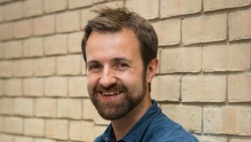
This year’s winner was Aroon Pressram, a masters student at the University of Florida, who presented a talk entitled “The hidden haemorrhage: visualizing brain bleeds”.
Pressram is developing a technique for rapid detection of brain bleeds in stroke patients. He explained that a patient presenting with stroke symptoms will typically be admitted to hospital for a CT scan, which involves injecting a contrast to aid visualization of the vessels in the brain. If a blockage is found, the patient receives revascularization therapy to restore the blood flow. But this treatment can actually put the patient at risk of developing a brain bleed or leakage of contrast into their brain. “That’s why it is essential that we perform follow-up imaging so we can identify the brain bleeds and reverse it,” he explained.
So how best to perform such follow-up imaging? MRI is accurate and provides high-quality images, but it is slow. CT scanning, meanwhile, is much faster but can’t distinguish brain bleeds from contrast in the brain. “There has to be a better way to get something accurate and something fast for the patient,” said Pressram. “Well there is. And It’s called dual-energy CT.”

Dual-energy CT works by performing two scans with different X-ray spectra, then combining the two data sets mathematically. The technique can separate signals due to a brain bleed from those coming from the contrast. Pressram notes that dual-energy CT is also more readily available than MRI and offers quicker scan times.
Following a literature review, Pressram realised that “we were the first people in the world doing research on this dual-energy scanner for stroke patients”. To investigate the application further, he assessed 500 stroke patients with dual-energy CT and found that the approach performed well in all cases, giving accurate results in a timely manner. “Healthcare professionals should be aware of this amazing technology that’s out there that can give them accurate results in a quicker time,” he concluded.
Improving prostate radiotherapy
Second place in the competition went to Ellie Bacon, a medical physics resident at the University of Nebraska Medical Center. Bacon described how a process called offline review could improve radiotherapy for prostate cancer patients.
Offline review – which Bacon referred to as “the single most important task that we do for our patients on a weekly basis” – involves examining the images taken during a patient’s treatment over the previous week to look for any potential errors that need to be quickly addressed and to follow tumour shrinkage over time.
For prostate cancer patients, one important parameter is how well they are able to fill their bladder from day to day. “We found that when patients are unable to fill their bladder 50% full for their treatment, they have a much higher chance of side effects such as bladder toxicity,” Bacon explained. “That got me thinking, is there a way that we can find these patients quickly so we can help them?”
Bacon proposed a simple addition to the offline review process, in which a patient’s bladder is categorized as “good” if it looks above 50% full, or “bad” for those below 50%. She performed a test in which her team assessed patients over three rounds, with additional visual clues provided each time: first, an outline of what a full bladder should look like from the patient’s original treatment plan; then an image of an empty bladder; and finally an estimate of what a 50% full bladder should look like.
“Each round, with more and more visual clues, they were able to quickly identify which patients were good or bad and needing our help,” said Bacon. “This confirmed my suspicion – we are able to quickly use offline review, which we already do for all of our patients, to identify the prostate cancer patients who need help.”
Once such patients are identified, their treatment plan can be adapted to better fit their bladder-fill average. This reduces their chance of side effects and improves quality-of-life following treatment. “The only question left is who else can we help with this offline review?” she concluded.
Keeping track of the tumour
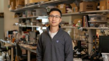
Taking third place in the MedPhys Slam, as well as winning the “people’s choice award” voted for by the audience, was Jason Luce, a PhD student at Loyola University. Luce told the attendees about an adaptive template-based tumour tracking algorithm for lung cancer radiotherapy.
Tumour tracking during radiotherapy is particularly important for patients with lung cancer. Breathing causes tumour motion, which leads to increased uncertainty in the tumour position. This requires the use of a larger treatment beam that can increase irradiation of healthy surrounding tissue. “But if you can actively track the tumour, you can use a more precise treatment beam, which means less radiation to healthy tissues,” Luce explained.
During image-based tracking, however, it’s possible to lose the tumour, particularly when using a large search window to cover all possible ranges of tumour motion. For example, Luce showed a case in which the tracking algorithm misidentified the location of the tumour as that of an extraneous image artefact.
He likened this tracking problem to that of looking for lost car keys. “Rather than search your entire house to find them, you can make your life easier by asking ‘where was the last place I saw them?’ In the kitchen? Just search that area, problem solved,” he said. “We’re taking that idea and applying it towards improving tumour tracking.”

MedPhys Slam highlights the art of science communication
The approach, Luce explained, involves finding the last place that the tumour was seen during tracking, and then reducing the search region to that area. He tested the technique on 229 X-ray images of a tumour in motion, performing tracking using an algorithm with a large search window, as well as one with a smaller adaptive search window.
The smaller adaptive search window provided a notable improvement in tumour tracking. With the static search window, about 12% of images exhibited poor tracking (significant differences between the actual and predicted tumour locations), while less than 1% were poorly tracked by the adaptive search window. “We’re improving tracking results and ideally improving patient care,” he said.
- SEO Powered Content & PR Distribution. Get Amplified Today.
- PlatoData.Network Vertical Generative Ai. Empower Yourself. Access Here.
- PlatoAiStream. Web3 Intelligence. Knowledge Amplified. Access Here.
- PlatoESG. Automotive / EVs, Carbon, CleanTech, Energy, Environment, Solar, Waste Management. Access Here.
- BlockOffsets. Modernizing Environmental Offset Ownership. Access Here.
- Source: https://physicsworld.com/a/brain-bleed-detection-study-wins-aapms-medphys-slam/
- :has
- :is
- 17
- 2018
- 2023
- 500
- 90
- a
- Able
- About
- above
- accurate
- actively
- actual
- actually
- adapted
- addition
- Additional
- admitted
- Aid
- algorithm
- All
- already
- also
- amazing
- an
- and
- annual
- any
- Application
- Applying
- approach
- ARE
- AREA
- areas
- Art
- artificial
- artificial intelligence
- AS
- assess
- assessed
- At
- attendees
- audience
- available
- average
- aware
- Bad
- based
- BE
- Beam
- below
- BEST
- Better
- between
- blood
- Brain
- breathing
- but
- by
- called
- CAN
- Cancer
- cancer patients
- car
- care
- case
- cases
- causes
- Center
- Chance
- Chapter
- choice
- click
- combining
- coming
- Communication
- competition
- Competitions
- competitors
- concluded
- CONFIRMED
- contrast
- could
- cover
- covered
- data
- data sets
- day
- described
- Detection
- developing
- differences
- different
- distinguish
- do
- doing
- due
- during
- each
- easier
- effects
- else
- Entire
- entitled
- Errors
- essential
- established
- estimate
- Examining
- example
- Explain
- explained
- FAST
- faster
- Feature
- fill
- Finally
- Find
- finding
- First
- fit
- florida
- flow
- follow
- following
- For
- found
- from
- full
- further
- get
- Give
- Giving
- good
- Have
- he
- healthy
- help
- her
- Hidden
- high-quality
- higher
- highlights
- Hospital
- House
- How
- However
- HTTPS
- i
- idea
- ideally
- identified
- identify
- if
- image
- images
- Imaging
- important
- improve
- improvement
- improves
- improving
- in
- Including
- Increase
- increased
- information
- Intelligence
- into
- investigate
- involves
- issue
- IT
- ITS
- jpg
- just
- keys
- large
- larger
- Last
- Leads
- left
- less
- Life
- like
- literature
- local
- location
- locations
- Look
- look like
- looking
- LOOKS
- lose
- lost
- make
- manner
- mathematically
- max-width
- me
- means
- Meanwhile
- medical
- Medical physics
- meeting
- methods
- minutes
- more
- most
- motion
- MRI
- much
- my
- Nebraska
- Need
- needing
- notable
- Notes
- now
- of
- Offers
- offline
- on
- ONE
- only
- open
- or
- organizers
- original
- our
- out
- outline
- over
- panel
- parameter
- part
- particularly
- patient
- patient care
- patients
- People
- perform
- performed
- performing
- Physics
- Physics World
- Place
- plan
- plato
- Plato Data Intelligence
- PlatoData
- poor
- Popular
- position
- possible
- potential
- precise
- preclinical
- predicted
- present
- Presentations
- presented
- previous
- Problem
- process
- professionals
- projects
- proposed
- provided
- provides
- put
- question
- quicker
- quickly
- Radiation
- Radiotherapy
- range
- rapid
- receives
- reduces
- reducing
- referred
- region
- requires
- research
- restore
- Results
- reverse
- review
- right
- Risk
- round
- rounds
- Said
- saw
- scan
- scanning
- scans
- Science
- Search
- seen
- selected
- separate
- session
- Sets
- she
- should
- showed
- side
- signals
- significance
- significant
- Simple
- single
- Slides
- slow
- smaller
- So
- something
- speakers
- Student
- Students
- Study
- such
- Surrounding
- Symptoms
- taken
- taking
- Talk
- Talks
- Task
- team
- Technology
- test
- tested
- than
- that
- The
- the world
- their
- Them
- then
- therapy
- There.
- These
- they
- Thinking
- Third
- this
- thompson
- those
- three
- thumbnail
- time
- times
- tissue
- tissues
- to
- took
- towards
- track
- Tracking
- treatment
- true
- two
- typically
- unable
- Uncertainty
- university
- use
- using
- vessels
- via
- visualization
- voted
- was
- Way..
- we
- week
- weekly
- WELL
- went
- were
- What
- when
- which
- while
- WHO
- why
- wide
- Wide range
- will
- window
- winner
- winners
- winning
- Wins
- with
- works
- world
- x-ray
- year
- you
- Your
- zephyrnet