- SEO Powered Content & PR Distribution. Get Amplified Today.
- PlatoData.Network Vertical Generative Ai. Empower Yourself. Access Here.
- PlatoAiStream. Web3 Intelligence. Knowledge Amplified. Access Here.
- PlatoESG. Carbon, CleanTech, Energy, Environment, Solar, Waste Management. Access Here.
- PlatoHealth. Biotech and Clinical Trials Intelligence. Access Here.
- Source: https://www.nanowerk.com/news2/gadget/newsid=64573.php
- :has
- :is
- :not
- 02
- 10
- 12
- 2%
- 2023
- 3d
- 4
- 5
- 6
- 7
- 8
- 9
- a
- Able
- accessible
- According
- achievement
- across
- added
- affected
- All
- allow
- almost
- already
- also
- Alzheimer’s
- an
- and
- any
- approach
- ARE
- arrangement
- AS
- aspect
- At
- Attempts
- BE
- because
- behind
- belonging
- between
- biology
- Brain
- brain cells
- brains
- broad
- but
- by
- CAN
- candidates
- cell
- Cells
- Center
- cerebral
- certain
- change
- Common
- communicate
- comparable
- components
- conditions
- Connections
- Connectivity
- control
- cortex
- could
- critical
- Date
- defined
- depth
- described
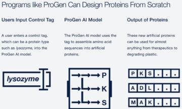
- Design
- developed
- Development
- developmental
- different
- disabilities
- Disease
- disorders
- do
- does
- down
- drug
- each
- easy
- employed
- enough
- equipment
- Even
- Every
- exactly
- explore
- field
- First
- Flexibility
- For
- form
- forming
- found
- from
- function
- functional
- further
- get
- Group
- Grow
- grown
- Growth
- had
- Have
- healthy
- help
- hold
- How
- HTTPS
- Hugely
- human
- Humans
- Imaging
- implications
- important
- improvements
- improving
- in
- inside
- instead
- interact
- interactions
- into
- IT
- journal
- jpg
- Keep
- lab
- Labs
- laid
- layer
- layers
- less
- like
- Limited
- Look
- looked
- major
- make
- MAKES
- many
- many people
- means
- mechanisms
- Media
- medium
- method
- methods
- miss
- model
- molecular
- more
- much
- neighboring
- network
- networks
- Neural
- neurodegenerative
- neurological
- neurology
- Neurons
- Neuroscience
- New
- next
- now
- of
- Offers
- often
- on
- On-Demand
- ONE
- operate
- operates
- or
- organization
- Other
- our
- over
- Oxygen
- Parkinson’s Disease
- parts
- past
- pencils
- People
- piece
- plato
- Plato Data Intelligence
- PlatoData
- potential
- powerful
- Precision
- pretty
- previous
- printing
- process
- produce
- Professor
- proper
- provides
- quite
- range
- reach
- refining
- relatively
- require
- researchers
- Results
- say
- says
- Scientist
- scientists
- send
- should
- signals
- situated
- Soft
- some
- speak
- special
- specialized
- specific
- specifically
- specificity
- stacking
- standard
- start
- stays
- Stem
- stem cells
- Still
- structure
- studied
- Study
- Studying
- success
- such
- support
- system
- Talk
- talking
- team
- technique
- techniques
- Testing
- than
- that
- The
- their
- Them
- themselves
- then
- they
- thin
- thing
- this
- though?
- Through
- time
- tissue
- tissues
- to
- together
- traditional
- treatments
- truly
- type
- types
- typical
- under
- underlying
- understand
- university
- us
- used
- using
- vertically
- very
- want
- was
- watching
- Way..
- we
- WELL
- went
- were
- What
- whatever
- when
- which
- with
- within
- working
- works
- would
- zephyrnet
- zhang
More from Nanowerk
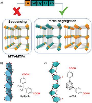
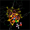
Engineering multi-metal MOFs with customizable properties
Source Node: 2387755
Time Stamp: Nov 17, 2023
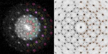
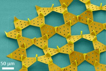
Nanoengineers create a quasicrystal from nanoparticles using DNA
Source Node: 2362566
Time Stamp: Nov 2, 2023
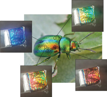
Beetles living in the dark teach us how to make sustainable colors
Source Node: 2513556
Time Stamp: Mar 13, 2024
Solar cell material can assist self-driving cars in the dark
Source Node: 2303751
Time Stamp: Sep 28, 2023
New structural insights could lead to mechanical enhancement in alloys
Source Node: 2494110
Time Stamp: Feb 23, 2024
Quantum mechanics could lead to stronger, more sustainable alloys
Source Node: 1976179
Time Stamp: Feb 24, 2023
Hummingbird beak points the way to designing complex micro machines
Source Node: 2216739
Time Stamp: Aug 16, 2023

New atomic-scale understanding of catalysis could unlock massive energy savings
Source Node: 2050160
Time Stamp: Apr 6, 2023
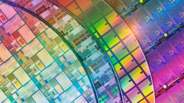
Accelerating sustainable semiconductors with ‘multielement ink’
Source Node: 2297397
Time Stamp: Sep 28, 2023