Light is critical for transmitting visual information to the brain; but light also impacts non-visual processes in the body, such as circadian rhythms, hormone secretion, pupil size and sleep cycles. Exposure to blue light is known to stimulate alertness and enhance cognitive performance, but the neural processes underlying this effect are not well understood. Now, researchers at the University of Liège in Belgium have used ultrahigh-field MRI to find out more about how light stimulates our brains, reporting their findings in Communications Biology.
Non-visual responses to light are mainly mediated by photosensitive retinal ganglion cells that express melanopsin, a photopigment that’s most sensitive to blue light at around 480 nm. These retinal neurons transfer light information to several areas of the brain associated with light-mediated behaviour. In particular, the pulvinar (a region of the posterior thalamus involved in attention control) is consistently activated in response to light, suggesting that the thalamus, a subcortical region, may play a key role in relaying non-visual light information to the cortex.
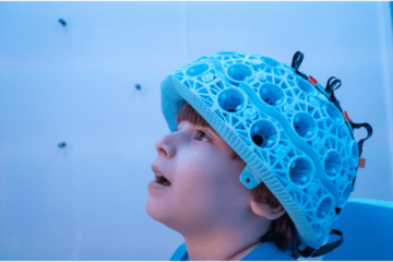
To investigate this hypothesis, first author Ilenia Paparella and colleagues in the GIGA-CRC laboratory used 7T functional MRI to record the brain activity of 19 healthy young participants while they completed an auditory oddball task known to elicit response in the posterior thalamus. During the task, in which random rare deviant tones were sounded amongst frequent standard tones, the volunteers were either in darkness or exposed to 30 s blocks of blue-enriched polychromatic or control orange light.
The researchers assessed how light exposure during the auditory task affected the connectivity from the thalamus to the intraparietal sulcus (IPS), an attentional-related area of the cortex. For each subject, they analysed changes in the blood oxygen level dependent (BOLD) signals from the thalamus and the IPS to infer their neuronal activity. They then used dynamic causal modelling to estimate the effective connectivity between the two brain regions.
The team’s analyses revealed that hearing a rare deviant tone during the auditory task excited both the pulvinar and the IPS in both hemispheres. Without light exposure, there was significant reciprocal negative influence between both regions.
The researchers found that only the blue-enriched light strengthened the connectivity from the posterior thalamus to the IPS, switching the influence of the posterior thalamus on the IPS from inhibition to excitation. “In other words, the active light modulated specifically the information flow from thalamic to cortical areas, while the control light, despite otherwise eliciting visual responses, was not affecting our network in any way,” they write.

High-resolution MRI enables direct imaging of neuronal activity
Importantly, the researchers computed two independent models for the left and right brain hemispheres, and saw that they yielded the same results. They also observed that the impact of blue-enriched light on connectivity correlated with changes in pupil size – another measure of the non-visual impact of light on physiology.
“To the best of our knowledge, our results provide the first empirical data supporting that blue wavelength light affects ongoing non-visual cognitive activity by modulating task-dependent information flow from subcortical to cortical areas,” the researchers write.
- SEO Powered Content & PR Distribution. Get Amplified Today.
- PlatoData.Network Vertical Generative Ai. Empower Yourself. Access Here.
- PlatoAiStream. Web3 Intelligence. Knowledge Amplified. Access Here.
- PlatoESG. Carbon, CleanTech, Energy, Environment, Solar, Waste Management. Access Here.
- PlatoHealth. Biotech and Clinical Trials Intelligence. Access Here.
- Source: https://physicsworld.com/a/ultrahigh-field-mri-reveals-how-blue-light-stimulates-the-brain/
- :is
- :not
- 160
- 19
- 25
- 30
- 7
- 90
- a
- About
- activated
- active
- activity
- affecting
- affects
- also
- amongst
- an
- analyses
- and
- Another
- any
- ARE
- AREA
- areas
- around
- AS
- assessed
- associated
- At
- attention
- author
- b
- Belgium
- BEST
- between
- Blocks
- blood
- Blue
- body
- bold
- both
- Brain
- Brain activity
- brains
- but
- by
- Cells
- Changes
- click
- cognitive
- colleagues
- Completed
- Connectivity
- consistently
- control
- correlated
- cortex
- course
- CRC
- critical
- cycles
- data
- de
- dependent
- Despite
- direct
- during
- dynamic
- each
- effect
- Effective
- either
- enables
- enhance
- estimate
- Ether (ETH)
- excited
- exposed
- Exposure
- express
- Find
- findings
- First
- flow
- For
- found
- frequent
- from
- functional
- Have
- healthy
- hearing
- hemispheres
- How
- HTTPS
- image
- Imaging
- Impact
- Impacts
- in
- independent
- influence
- information
- investigate
- involved
- issue
- jpg
- Key
- knowledge
- known
- left
- Level
- light
- mainly
- max-width
- May..
- measure
- min
- modelling
- models
- more
- most
- MRI
- Nature
- negative
- network
- Neural
- neuronal
- Neurons
- now
- observed
- of
- on
- ongoing
- only
- open
- or
- Orange
- Other
- otherwise
- our
- out
- Oxygen
- participants
- particular
- performance
- Physics
- Physics World
- plato
- Plato Data Intelligence
- PlatoData
- Play
- Point
- processes
- provide
- random
- RARE
- record
- recording
- region
- regions
- Reporting
- researchers
- response
- responses
- Results
- Revealed
- Reveals
- right
- Role
- s
- same
- saw
- scans
- sensitive
- several
- signals
- significant
- Size
- sleep
- sounded
- specifically
- standard
- stimulates
- subject
- such
- Supporting
- T
- Task
- that
- The
- the information
- their
- then
- There.
- These
- they
- this
- thumbnail
- time
- to
- TONE
- transfer
- true
- two
- underlying
- understood
- underwent
- university
- used
- volunteers
- was
- Way..
- WELL
- were
- which
- while
- with
- without
- words
- world
- write
- yielded
- young
- zephyrnet